Electrocardiogram (ECG): It’s Indications, and How to Read the Report
Last Updated on 04/01/2026 by Helal Medical
The heart is one of the most vital organs in the body. Monitoring its functions is crucial for maintaining overall health. One of the most common and essential tests to evaluate heart activity is the electrocardiogram (ECG).
Electrocardiogram (ECG) Key points
- Intro: What is an ECG?
- Section 1: Why do you need one?
- Section 2: ECG types
- Section 3: How to Prepare for an ECG
- Section 4: Understanding the Report. How to read the waves (P, QRS, T) – This is your high-value technical content.
- Section 5: FAQ. Use the FAQ schema we discussed earlier.
What is an ECG?
It is a test to record the electrical signals of the heart. It helps doctors to know rhythm problems.and used to detect heart disease and other cardiac issues.
You are a medical student trying to understand how an ECG works or a patient preparing to undergo the test. Hence, this article will help you. It provides a clear overview of ECG. It covers the machines used, when doctors recommend it, and how to interpret the results.
What is an Electrocardiogram (ECG) Machine?
It’s a device that records the heart’s electrical activity through electrodes attached to the skin. These electrodes pick up tiny electrical impulses generated each time the heart beats. The signals are then amplified and displayed as waveforms on paper or a screen.
Modern ECG machines vary in size and functionality:
- Standard 12-lead ECG machines are commonly used in hospitals and clinics. They record electrical activity from 12 different angles, providing a detailed picture of the heart.
- Portable/handheld ECG devices are useful for quick assessments, often used in emergency settings or by patients at home.
- Holter monitors are small wearable ECG devices that record heart activity over 24–48 hours. They are useful for detecting irregular rhythms that can’t be detected during a short test.
- Stress test ECG machines that can record the heart’s activity while the patient exercises. They give valuable information about blood flow. They also assess heart functions under stress.
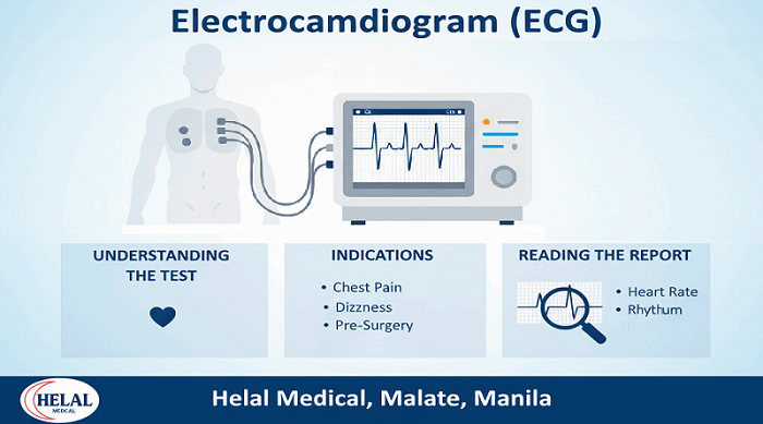
Indications for Doing an ECG
Doctors recommend an ECG for a variety of reasons. It is a quick, painless, and non-invasive test, making it a first-line investigation in many situations. Common indications to do an ECG include:
- Chest pain. To rule out angina or a heart attack.
- Palpitations or irregular heartbeat. To detect arrhythmias like atrial fibrillation, tachycardia, or bradycardia.
- Shortness of breath. To assess whether heart problems are contributing to breathing difficulty.
- High blood pressure. Long-term hypertension can damage the heart, and an ECG helps detect related issues.
- Dizziness or fainting (syncope). Sometimes caused by irregular heart rhythms.
- Routine screening. Especially before surgery, in annual checkups for older adults, or for athletes to assess cardiac risk.
- Monitoring heart conditions. For patients with a history of heart attack, pacemaker implantation, or ongoing cardiac treatment.
In emergency medicine, ECG is a vital tool as it can quickly detect life-threatening heart problems. These include myocardial infarction (heart attack) or severe arrhythmias. Read more about: Heart Attack Prevention.
Types of ECG Tests
Depending on your symptoms, your doctor may recommend:
- Resting ECG: done while you’re lying down. Most common test to check heart rhythm and structure.
- Exercise ECG (Stress Test): performed while walking on a treadmill; evaluates how your heart responds to physical activity.
- Holter Monitor: a portable ECG worn for 24–48 hours to track heart rhythm over time.
How to Prepare for an ECG
- Wear comfortable clothes; avoid lotions or oils on your chest.
- Inform your doctor about any medications you’re taking.
- Relax and breathe normally during the test — it’s quick and painless.
No fasting or special preparation is required.
Reading the ECG Report
For many patients, the Electrocardiogram (ECG) paper looks like a confusing set of spikes and waves. However, each part of the tracing corresponds to a specific activity of the heart. Understanding these basics can help both students and patients appreciate what doctors are looking at.
1. The ECG Waves
- P wave:
It shows atrial depolarization (the electrical activity that makes the atria contract). - QRS complex
It’s the ventricular depolarization (the main pumping chambers contracting). - T wave
It’s the ventricular repolarization (the recovery phase before the next beat).
2. Heart Rate and Rhythm
By measuring the spacing between QRS complexes, doctors can decide the heart rate and whether it is regular or irregular. A normal heart rate is between 60–100 beats per minute.
3. Intervals and Segments
- PR interval:
Shows the time taken for electrical signals to move from the atria to the ventricles. - ST segment:
Very important in detecting heart attacks. Elevation or depression of this segment indicates reduced blood supply to the heart muscle. - QT interval:
Represents the total time of electrical activity in the ventricles. Abnormalities can predispose to dangerous arrhythmias.
4. Common Abnormalities Detected on ECG
- Arrhythmias – Irregular heartbeats like atrial fibrillation, atrial flutter, or ventricular tachycardia.
- Heart attack (myocardial infarction) – Shown by changes in the ST segment or T waves.
- Heart enlargement (hypertrophy) – Abnormally tall waves may suggest thickened heart muscle.
- Electrolyte imbalances – Low potassium, calcium, or magnesium can cause distinctive ECG changes.
- Drug effects – Some medications influence ECG patterns, as digoxin or anti-arrhythmic drugs.
5. Limitations of ECG
While ECG is highly useful, it is not a perfect test. Some heart conditions may not show up on a resting ECG. This is why doctors order extra tests as echocardiography, cardiac MRI, or stress testing.
Summary
The electrocardiogram (ECG) is a powerful yet simple diagnostic tool that provides valuable information about the heart’s electrical activity. With just a few electrodes placed on the chest and limbs, doctors can detect rhythm problems. They can also identify past or ongoing heart attacks and many other cardiac conditions.
- ECG machines come in various types, from standard 12-lead models to portable devices and long-term monitoring tools.
- Indications for ECG include chest pain, palpitations, dizziness, fainting, shortness of breath, high blood pressure, and routine health checks.
- Reading an ECG report involves analyzing the P wave, QRS complex, and T wave. It also includes measuring intervals. Additionally, one looks for abnormalities in rhythm or ST segments.
For patients, it’s reassuring to know that an ECG is quick, painless, and risk-free. For healthcare professionals, it remains one of the most indispensable tools in diagnosing and managing heart disease.
Understanding ECG basics empowers patients to better engage in discussions with their doctors. It also helps medical students build a strong foundation in cardiology. Cardiovascular diseases remain the leading cause of death globally. In this context, the humble ECG continues to save lives every day.
If you suspect that you may have symptoms, Helal Medical can help, offering quick, private, and convenient testing options. You may contact us here: Facebook page.
Read More About Heart Attack:
- Heart attack, can be spotted by a rapid low cost by blood test?
- Electrocardiogram (ECG): It’s Indications, and How to Read the Report
- Heart Health: How to Prevent Heart Disease Before It Starts
- Risk for Heart Disease: Don’t Let High Cholesterol Put You at Risk
Read More About ECG:
- ECG: Top 5 Reasons Your Doctor May Recommend It
- ECG Test in Manila: What to Expect, Cost, and Benefits
- ECG vs. Echocardiogram: Which Test Do You Need?
- Electrocardiogram (ECG): It’s Indications, and How to Read the Report
This article provides general information. Always consult with a healthcare professional for personalized medical advice.
Discover more from Helal Medical Manila
Subscribe to get the latest posts sent to your email.


